Imaging Based Phenotyping

For imaging-based phenotyping, EPIC provides access to two different modalities, namely to an ultrasound/photoacoustic system and a computer tomograph.
High resolution micro-ultrasound and photoacoustic imaging system for real-time visualization
The system allows multiple applications such as anatomical, functional and physiological analyses, image-guided techniques, molecular and contrast-based imaging as well as photoacoustic imaging.
Ultrasound Imaging
Using the ultrasound capability of the system, applications inlcude but are not limited to cardiac imaging, vascular imaging, tumor imaging, and molecular imaging. The system supports the following imaging modes: B-Mode, M-Mode, Power Doppler Mode, Pulsed Wave Doppler Mode, Color Doppler Mode, PW Tissue Doppler Mode and Contrast Mode. Measurements of the study data can be performed in all modes and include applications specific calculations for cardiology, abdominal anatomy, tumor analysis, embryology and vascular anatomy.
Photoacoustic Imaging
The photoacoustic imaging is a new in-vivo hybrid imaging modality that combines the sensitivity and contrast of optical imaging with the depth and resolution of ultrasound. When pulsed laser light illuminates tissue, the optical absorbers there (such as hemoglobin) undergo thermoelastic expansion, generating an acoustic pressure wave which is detected with an ultrasound transducer.
The Vevo LAZR system incorporates photoacoustic imaging into high-resolution ultrasound. The ultrasound imaging provides a high-resolution frame-of-reference for identifying anatomy, while the photoacoustic imaging enables functional measurements such as oxygen saturation, total hemoglobin and the microdistribution of biomarkers. As such, the system provides optical contrast for blood and molecular imaging, high-resolution at depth, real-time non-invasive anatomical, functional and molecular data.
Available device: Vevo® 2100 with Vevo LAZR imaging system
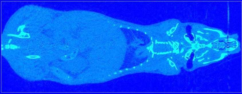
In-vivo micro-computed tomography (CT)
Micro-CT is based on X-Rays. Up to 1000 projection images are taken over 180° and reconstructed to a 3D image. Micro-CT can be used to image tissues with different X-ray absorption coefficients. It is very well suited to image bone and other calcified tissues.
Available device: VivaCT40 (SCANCO Medical)
Common CT applications:
Bone imgaing
- Bones can be imaged as whole or just certain areas. Bones can be imaged at a resolution of up to 10 um voxel size.
- The scanner offers sophisticated software for the analysis of bone tissue. .
Vascular imaging
- Using blood pool contrast agents it is possible to visualize and quantify larger blood vessel (min ca. 50 um diameter).
Whole body imaging:
- Images of a whole body can be used as diagnostic tool, e.g. to find tumors, fractures etc. Also body fat content can be determined.
Lung imaging:
- Imaging of the lung requires gating. This means, images are only taken at a certain phase in the respiratory cycle.
- It is also possible to quantify tumors or non-ventilated areas (e.g. lung fibrosis).